A team of investigators based at Illinois has been awarded an $8 million grant over five years to study DNA structure as part of the National Institutes of Health Common Fund’s 4D Nucleome Program.
For years genome-mapping technology has understood DNA as a linear code, but that’s not how it exists in the cell: it’s tangled up in 3D inside the nucleus, with higher- and lower-density areas. It’s estimated that each human cell contains approximately two meters of DNA, squeezed inside the cell’s microscopic nucleus.
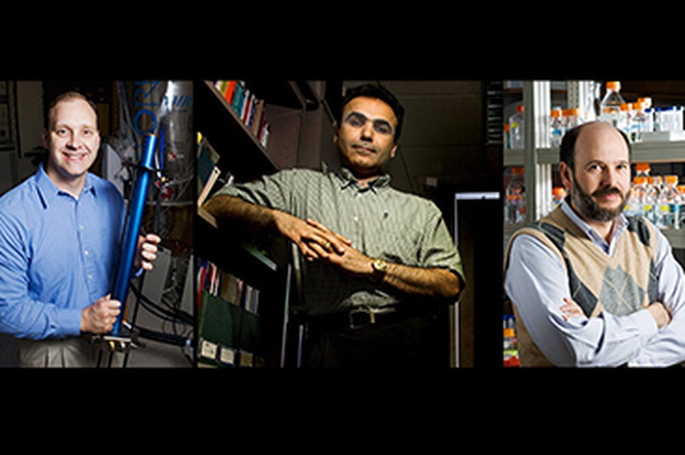
Moreover, DNA exists in the fourth dimension as well—gene clusters can shift their positions over time. Professor of cell and developmental biology Andrew Belmont is leading a team from around the world to study the implications of this positioning on development and disease.
“Understanding nuclear structure at this level is an important first step,” Belmont said.
Chromosomal DNA isn’t homogenously compacted or straight. It can fold and coil around itself, with non-coding and inactive regions compacted several thousand-fold and more active regions still compacted hundreds to thousands-fold. At the same time, different chromosome regions are positioned in different parts of the nucleus.
“Certainly there’s a strong correlation between compaction and function,” Belmont explained. “Things that are less folded replicate earlier, and are more active in transcription. But at the same time, particularly in mammalian cells, there’s an increased density of genes lying together on the DNA in certain chromosome bands and a markedly decreased density in other bands.”
These gene-rich areas tend to cluster near nuclear speckles, small objects located toward the center of the nucleus, or other regions in the nuclear interior, while gene-poor regions are often located near the nuclear periphery.
“Adding to the complexity, these positions change when gene activity changes. We don’t really know why genes are ordered along the chromosome DNA sequence and why chromosomes are specifically positioned within the nucleus these ways, but it’s very striking,” Belmont said.
Measuring exactly where different parts of the genome are located within the nucleus is a major goal of Belmont’s team. The group headed by Belmont is one of six 4D Nucleome Center grants studying organization and function within the nucleus.
Another 23 awarded grants span several 4D Nucleome funding initiatives, including developing new tools for genome mapping or imaging of chromosomes within the nucleus, understanding nuclear bodies, and managing the massive amount of data the 4D Nucleome Program will produce.
Belmont’s project, titled “Combined cytological, genomic, and functional mapping of nuclear genome organization” includes four additional investigators: Jian Ma, professor of bioengineering and Huimin Zhao, professor of chemical and biomolecular engineering, both at Illinois; David Gilbert at Florida State University; and Bas van Steensel at the Netherlands Cancer Institute. Belmont, Ma, and Zhao are also members of the Carl R. Woese Institute for Genomic Biology at Illinois.
Bas van Steensel invented a tagging method that adds a distinctive mark to DNA in direct contact with the nuclear wall, while Belmont’s own research group has developed a complementary method, which uses antibody staining to measure the cytological distance between DNA and the stained nuclear compartment.
Combining these two methods has created verifiable location maps for genes within the nucleus, Belmont said.
“Now the idea is to label different structures within the nucleus, generating contact frequency and distance maps for all the chromosomes…eventually, our goal is to come up with machine-learning computer algorithms that will take all these data and better predict where chromosomes are in the nucleus,” Belmont said.
This algorithm and data mining approach will be the focus of Ma’s group, whose goal is to use this modeling to better predict chromosome position and trajectories within the nucleus.
A second goal of Belmont’s team is to overlay this structural map with a complementary functional genomic map. Ma’s group will use a computational model to assemble replication-timing data from the Gilbert group and reporter-gene activity from the van Steensel group, together with other genomic data, to correlate DNA sequence and nuclear position with gene function.
By inserting large DNA pieces synthesized by Zhao’s group, Belmont’s team will “rewire” chromosome regions to different nuclear locations and then determine the functional consequences of this rewiring.
Belmont believes this research, together with other 4D Nucleome projects, will dramatically further our understanding of the connections between nuclear organization and genomic function.