
Capturing action on camera is difficult. Just ask any sports photographer who has tried to capture a frozen moment in football. But trying to take action shots on the microscopic level, with atom-by-atom detail, has been beyond the current technology—until now. U or I researchers, working with scientists in Germany, have found a way to take the first detailed picture of a ribosome in action.
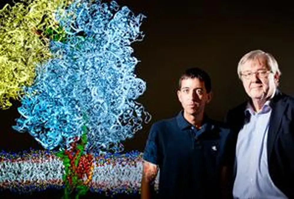
“We were able to get the first snapshot ever that sees a newly born protein moving out of a ribosome and into a SecY channel and then into the cell’s membrane,” says Klaus Schulten, U of I professor of physics and chemistry. What’s more, this picture is a detailed action shot, depicting the roughly 3 million atoms that make up the ribosome and other players on the microscopic field.
According to Schulten, ribosomes are found in every living cell, and they carry out a central function—to read genetic information for the synthesis of protein. Ribosomes have also become a hot topic in scientific circles in light of the 2010 Nobel Prize for chemistry, which went to three scientists who solved the structure of the ribosome. The U of I work builds upon this award-winning research.
“The ribosome is clearly on the minds of many biologists and biophysicists because it’s a key component of living cells,” Schulten says, adding that ribosomes also play an important role in the development of antibiotics. Therefore, seeing them in action has important medical benefits.
The 2010 Nobel laureates determined the structure of a ribosome in crystallized form. “But in this form,” he says, “the ribosomes all stand at attention and don’t do very much because they are closely packed together.”
Schulten says it’s like taking a photograph of football players lined up on the sidelines singing the national anthem. “You can see the players, but if you want to teach someone how the players function on the field, a photograph of them on the sidelines doesn’t tell you very much. You need to take a picture of the players in action.”
So Schulten and physics graduate student James Gumbart set out to do just that. They worked with Roland Beckmann at the University of Munich, who has been able to “shock-freeze” the action of a ribosome and capture the image with electron microscopy.
The problem is that the images are fuzzy.
“Electron microscopy doesn’t see the detail,” Schulten points out. Going back to the analogy of the football photographer, he says it’s like a blurry photograph in which you might be able to make out the player’s helmet, torso, and legs, but not much more. “You don’t see the player’s eyes, nose, or fingers, and what you want is a detailed look—to see every knuckle on their fingers.”
Schulten and Gumbart’s task was to bring these fuzzy images into focus by filling in the details with what they call a “computational microscope.” The computational microscope is actually a computer program developed at the U of I over the past two decades for close to $20 million. It uses data to fill in details that an electron microscope misses.
To image the ribosome’s interaction with the membrane, Beckmann’s team used small disks of membrane held together with belts of engineered lipoproteins. University of Illinois biochemistry professor Stephen Sligar developed and pioneered the use of these “nanodiscs.”
In Schulten and Gumbart’s work, their computational microscope took the Nobel laureates’ data on ribosome structure and used it to fill in details of Beckmann’s image of the ribosome in action.
The result was a groundbreaking image of a newly born protein emerging from a ribosome and entering the cell membrane through the SecY channel. The image sheds new light on the process, revealing how a “signal sequence” at the end of a growing protein affixes itself to the outside of the channel and begins the process of threading a protein into the cell membrane.
Schulten says that the ribosome is a key drug target for antibiotics, making ribosome studies extremely important in the ongoing war against bacterial infections. His team now hopes to create a series of snapshots of the ribosome in action, essentially creating a movie of the process. Among other things, this would enable them to see how antibiotics interfere with processes inside bacteria.
“Understanding the ribosome better and how an antibiotic interferes with it is very relevant,” he says. “It may be one of the most important things we can do right now in medicine.”


