
A geologist, a microscopist and a doctor walk into a lab and, with their colleagues from across the nation, make a discovery that overturns centuries of thought about the nature and composition of kidney stones. The team’s key insight, reported in the journal Scientific Reports, is that kidney stones are built up in calcium-rich layers that resemble other mineralizations in nature, such as those forming coral reefs or arising in hot springs, Roman aqueducts or subsurface oil fields.
Most importantly for human health, the researchers found, kidney stones partially dissolve and regrow again and again as they form.
This contradicts the widely held notion that kidney stones are homogenous rocks that never dissolve and are different from all other rocks in nature, said University of Illinois geology and microbiology professor Bruce Fouke, who led the new research with Jessica Saw, a medical student at the Mayo Clinic School of Medicine and PhD student at the U of I; and Mayandi Sivaguru, an associate director of the Carl Zeiss Laboratories@Location at the Carl R. Woese Institute for Genomic Biology at the U of I.
“Contrary to what doctors learn in their medical training, we found that kidney stones undergo a dynamic process of growing and dissolving, growing and dissolving,” Fouke said. “This means that one day we may be able to intervene to fully dissolve the stones right in the patient’s kidney, something most doctors today would say is impossible.
“Instead of being worthless crystalline lumps, kidney stones are a minute-by-minute record of the health and functioning of a person’s kidney,” he said.
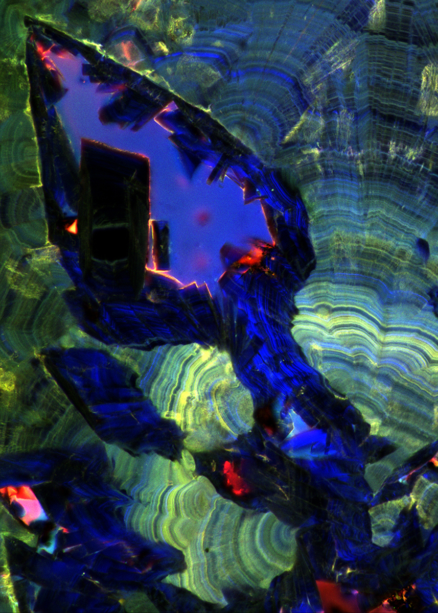
The findings were the result of looking at kidney stones much more closely and with a broader array of light and electron microscopy techniques than others had employed before, said Sivaguru, the lead author of the study who led the microscopy work. The methods included bright-field, phase-contrast, polarization, confocal, fluorescence and electron microscopy, with newly invented combinations of these tools and X-ray spectroscopy.
Many of the techniques are commonly employed in geology and geobiology (for example, Fouke has studied hot springs, coral reefs, and how growing limestone deposits in the Roman aqueducts affected water flow to the city in ancient times) but have not been used to study mineralizations in living organisms, like the kidney stones and gallstones that form in the human body, Fouke said. In particular, the use of ultraviolet light, which causes some minerals and proteins to fluoresce at different wavelengths, offered a vast new treasure trove of information.
A recently developed technology, Airyscan super-resolution microscopy, allowed the team to view the samples at 140-nanometer resolution, a much higher magnification than is normally possible with light microscopy.
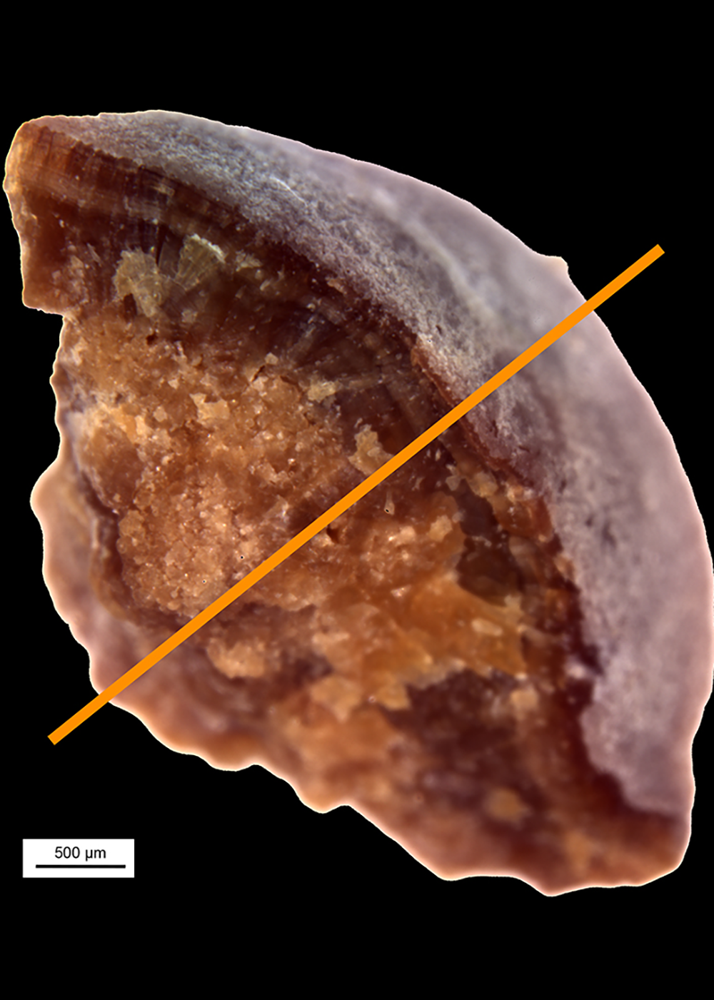
The effort resulted in spectacularly clear, colorful images of the interior growth history of the kidney stones, revealing that they are built up in alternating thin layers of organic matter and crystals, interrupted in places with jutting interior crystals.
In the earliest stages of kidney stone development, the researchers found, crystals of a hydrated form of calcium oxalate adhere to one another, forming a big, irregular clump. Layers of organic matter and crystals build up on top of this inner core, creating an outer shell. The stones continue to dissolve and grow. Being able to see the layers clearly for the first time made it possible to recreate this geological history, Fouke said.
“In geology, when you see layers, that means that something older is underneath something younger,” he said. “One layer may be deposited over the course of very short to very long periods of time.”
But many of the layers were disrupted, revealing that part of the stones – usually the interior dihydrate crystals – had dissolved. New crystals of a dehydrated form of calcium oxalate had begun to grow again within those voids.
“Therefore, just one rock represents a whole series of events over time that are critical to deciphering the history of kidney stone disease,” Fouke said.
Researchers and doctors who study and treat kidney disease will now need to rethink their basic assumptions, Saw said.
“Before this study, it was thought that a kidney stone is just a simple crystal that gets bigger over time,” she said. “What we’re seeing here is that it’s dynamic. The stone is growing and dissolving, growing and dissolving. It’s very rich with many components. It’s very much alive.”
Fouke, Saw and Sivaguru are affiliates of the Carl R. Woese Institute for Genomic Biology. Saw is pursuing a doctoral degree in molecular and integrative physiology at Illinois.
The Mayo Clinic and University of Illinois Strategic Alliance for Technology-Based Healthcare, the Mayo Clinic O’Brien Urology Research Center and the National Aeronautics and Space Administration Astrobiology Institute supported this research.